

Fetal brain atlas
A normative atlas of fetal brain maturation from 3D ultrasound, built from the INTERGROWTH-21st study, with labelled brain structures and growth curves. Explore it below. The files are available from the INTERGROWTH-21st website.
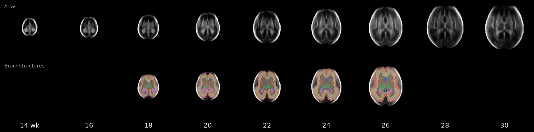
Drag to move through the brain · scroll to change slice · right-drag to adjust contrast · in 3D, drag to rotate
Growth curves
Normative growth of 28 brain measures from 18 to 27 weeks' gestation (structure and lobe volumes, cortical surface area, depth and thickness, and Sylvian fissure depth), from 4,205 3D ultrasound scans in the INTERGROWTH-21st study (Wyburd et al., 2026). The band shows the 3rd to 97th centile and the line the median.
Measures are for one hemisphere (the side further from the probe), except total brain and cavum septum pellucidum. Curves are from the published equations (Supplementary Table 8). For academic research use only; not a clinical tool and not for commercial use.
Download and cite
Brain structures and centiles
Label maps aligned to the atlas (10 structures and 5 cortical lobes), tables of label names and colours, and growth centiles by week.
Label and measure new scans
The OMNI ultrasound toolkit runs the same pipeline used in the paper: align a 3D scan to the atlas, label the brain structures, compute volumes and compare them with the growth curves.
The official copies of the atlas and labels, with the full terms of use, are on the INTERGROWTH-21st website.
Free for academic use. Commercial use is not permitted. Please cite the papers below (licence: CC BY-NC 4.0).
References
- Namburete AIL, Papież BW, Fernandes M, Wyburd MK, Hesse LS, Moser FA, et al. Normative spatiotemporal fetal brain maturation with satisfactory development at 2 years. Nature 623, 106–114 (2023). https://doi.org/10.1038/s41586-023-06630-3
- Wyburd MK, Kennedy SH, Fernandes M, Dinsdale NK, Hesse LS, Gunier RB, et al. Normative growth trajectories of fetal brain regions validated by satisfactory maturation of neurodevelopmental domains at 2 years of age. Nature Communications 17, 3073 (2026). https://doi.org/10.1038/s41467-026-69657-w